08 · At work
The finished protein at work: haemoglobin
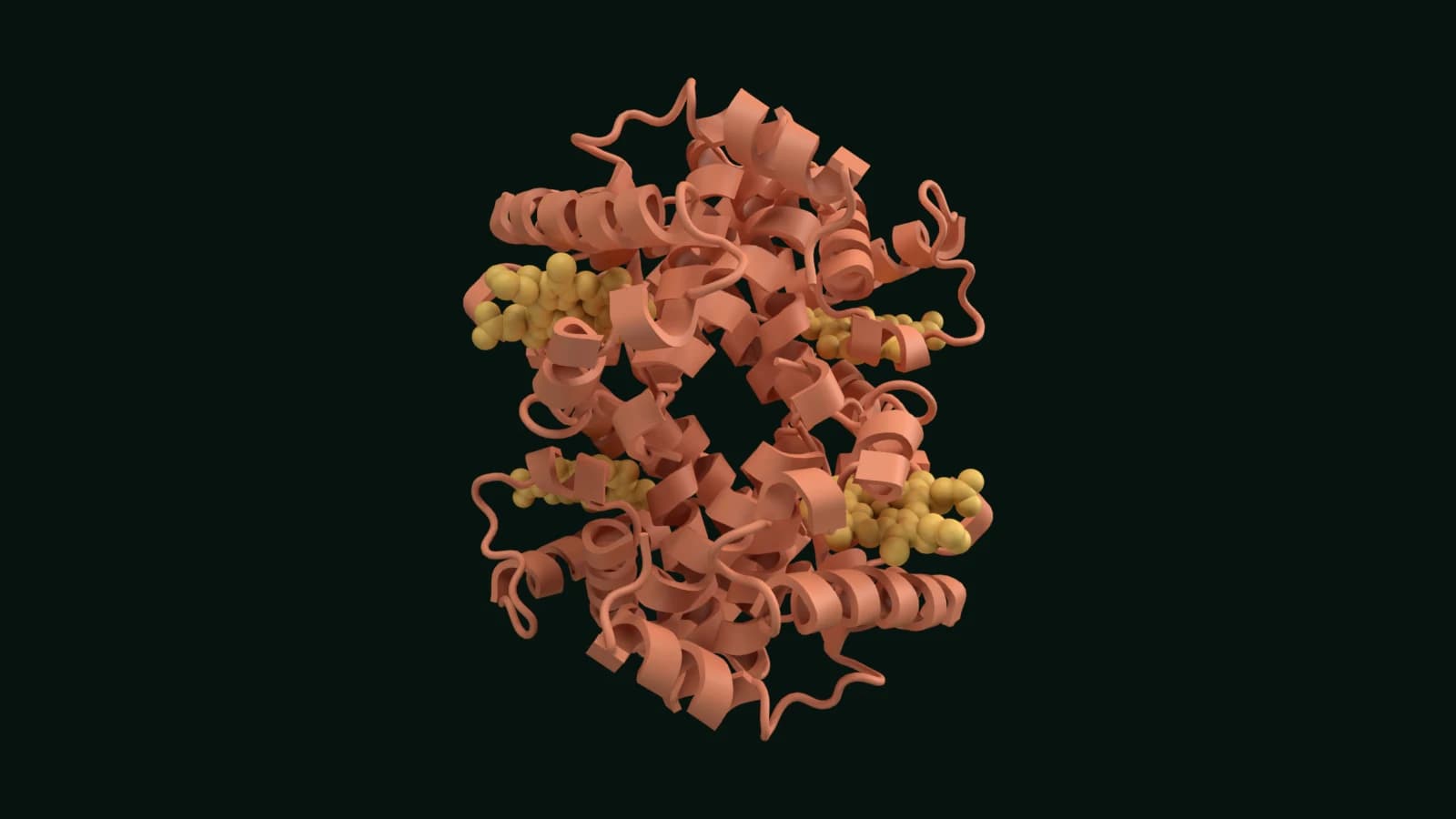
A folded protein can now do its job. Inside red blood cells, haemoglobin collects oxygen as blood passes through the lungs and hands it over to the tissues. Each molecule is built from two α and two β chains. Every chain holds a haem, a flat ring with an iron atom at its centre, and each iron can bind one oxygen molecule.
The four sites cooperate. When one binds oxygen the protein shifts shape and the others bind more easily, so haemoglobin loads up fully in the lungs and gives its oxygen up where oxygen is scarce. This model is human oxyhaemoglobin at 1.25 Å resolution, with oxygen on all four haems.
What you are looking at
- The α and β chains, two of each, each wrapped around its own haem.
- Four haem groups, each with an oxygen molecule bound to its iron.
- Structure
- Human oxyhaemoglobin. PDB 2DN1 (opens the RCSB PDB in a new tab)
- Method
- X-ray crystallography, 1.25 Å, released 2006
- Size
- About 7 nm across, roughly a fifth of the width of the ribosome.
- Paper
- Park SY, Yokoyama T, Shibayama N, et al. (2006). 1.25 Å resolution crystal structures of human haemoglobin in the oxy, deoxy and carbonmonoxy forms. J Mol Biol 360:690-701. doi:10.1016/j.jmb.2006.05.036
- Chromatin12 nm
- Transcription17 nm
- Splicing36 nm
- Export45 nm
- Translation32 nm
- Exit tunnel32 nm
- Folding17 nm
- At work7 nm
Sizes are in nanometres; 1 nm is a millionth of a millimetre. The eight structures come from separate experiments published between 2006 and 2025, so they show the stages of making a protein, not one molecule passing through them. All the proteins are human. The RNA being spliced in step 3 is listed as coming from an adenovirus, and in step 5 the message is synthetic and the tRNAs have a bacterial sequence.