Skip to content
MBiotech.
All tools

Educational tool

Inside a cell

A guided 3D tour of how a human cell turns a gene into a working protein, in eight real structures: chromatin, transcription, splicing, export, the ribosome, the exit tunnel, a folding chaperonin and haemoglobin at work.

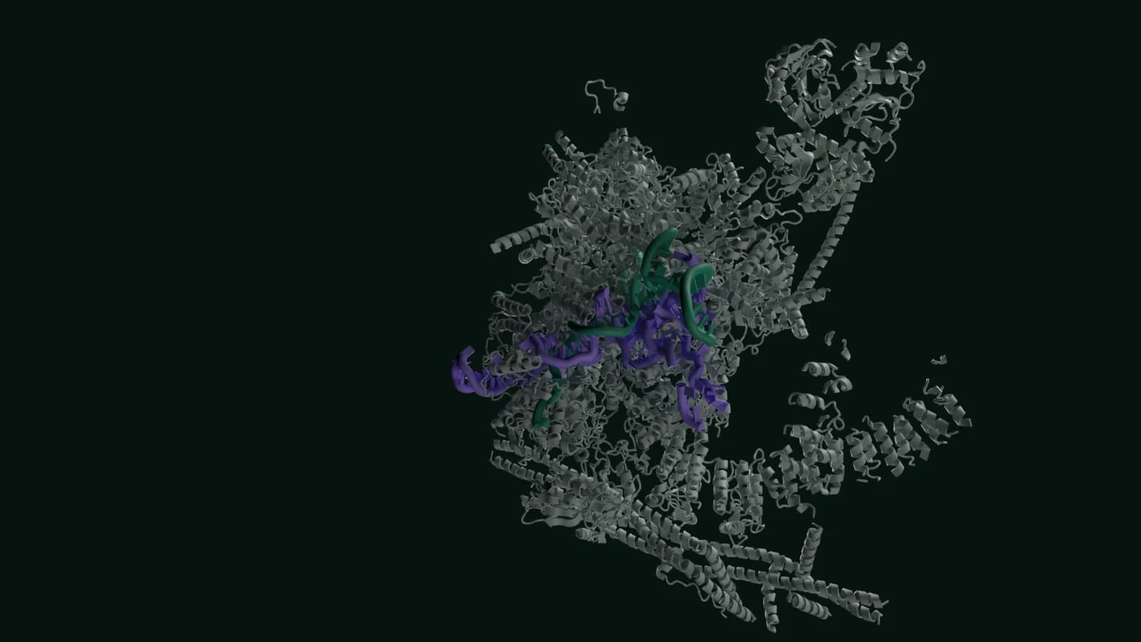
Human post-catalytic spliceosome, PDB 6QDV: a large grey protein complex, partly cut away, with violet small nuclear RNAs and green messenger RNA at its centre.

Mol* viewer, about 1.5 MB

  • Ribosomal and small nuclear RNA
  • Messenger RNA
  • Intron (cut out)
  • Protein machinery

3 / 8

03 · Splicing

The spliceosome removes an intron

A human gene is usually interrupted: stretches that are kept in the finished messenger RNA (exons), which between them carry the protein-coding sequence, alternate with stretches that are not (introns). The first RNA transcript contains both, so the introns have to be cut out. The spliceosome, a large machine made of proteins and small nuclear RNAs, removes each intron and joins the exons on either side.

This human spliceosome was caught just after it joined two exons. The cut-out intron is still held, as a loop called a lariat. The RNA being spliced, called MINX, is listed in the PDB entry as coming from human adenovirus 2.

What you are looking at

  • The U2, U5 and U6 small nuclear RNAs. RNA, not protein, carries out the splicing chemistry: U6 positions the metal ions at the active site.
  • The two joined exons: 14 nucleotides of them are resolved.
  • The cut-out intron, held as a lariat: 42 of its nucleotides are resolved.
  • 49 protein chains, among them PRPF8, a scaffold that positions the small nuclear RNAs, and the exon junction complex, which splicing leaves on the mRNA.
Structure
Human post-catalytic spliceosome (P complex). PDB 6QDV (opens the RCSB PDB in a new tab)
Method
Cryo-EM, 3.3 Å, released 2019
Size
About 36 nm across. Human spliceosome; the RNA it is splicing is listed as coming from an adenovirus.
Paper
Fica SM, Oubridge C, Wilkinson ME, et al. (2019). A human postcatalytic spliceosome structure reveals essential roles of metazoan factors for exon ligation. Science 363:710-714. doi:10.1126/science.aaw5569
On one scale
  • Chromatin12 nm
  • Transcription17 nm
  • Splicing36 nm
  • Export45 nm
  • Translation32 nm
  • Exit tunnel32 nm
  • Folding17 nm
  • At work7 nm

Sizes are in nanometres; 1 nm is a millionth of a millimetre. The eight structures come from separate experiments published between 2006 and 2025, so they show the stages of making a protein, not one molecule passing through them. All the proteins are human. The RNA being spliced in step 3 is listed as coming from an adenovirus, and in step 5 the message is synthetic and the tRNAs have a bacterial sequence.

The whole tour as text

Every step's explanation, structure and paper on one page, to read straight through, search or print.

Read all eight steps
  1. 01 · Chromatin

    A gene, packed into chromatin

    Most cells in your body hold two copies of your genome, one from each parent, each about 3.1 billion base pairs long. That DNA is not left loose: it is wound around histone proteins into nucleosomes, which compact it and limit which parts the cell's machinery can reach.

    This human nucleosome was solved by X-ray crystallography. Its 146 base pairs of DNA wrap about 1.65 times (a little under two turns) around a core of eight histones, two each of H2A, H2B, H3 and H4. The grip is a balance: tight enough to keep the DNA packed and protected, loose enough for the enzymes that read genes to get in.

    What you are looking at

    • The DNA double helix: 146 base pairs wrapped about 1.65 times around the core.
    • The eight histones. Their tails reach outwards; much of their length is too disordered to be seen in the crystal.
    Structure
    Human nucleosome core particle. PDB 3AFA (opens the RCSB PDB in a new tab)
    Method
    X-ray crystallography, 2.5 Å, released 2010
    Size
    About 12 nm across at its widest and about 6.5 nm thick.
    Paper
    Tachiwana H, Kagawa W, Osakabe A, et al. (2010). Structural basis of instability of the nucleosome containing a testis-specific histone variant, human H3T. Proc Natl Acad Sci U S A 107:10454-10459. doi:10.1073/pnas.1003064107
    Show step 1 in the tour
  2. 02 · Transcription

    RNA polymerase II copies the gene into RNA

    To read a gene, RNA polymerase II separates the two DNA strands and reads one of them, the template strand, building an RNA that base-pairs with it. The RNA therefore has the same sequence as the other (coding) strand, with U in place of T. It is built one nucleotide at a time at its growing end.

    Researchers pulled these polymerases out of human cell nuclei while they were transcribing the cell's own DNA, then imaged them by cryo-electron microscopy. In this model, 11 nucleotides of new RNA are resolved, and 10 of them are still paired with the template strand inside the enzyme.

    What you are looking at

    • The DNA the enzyme is moving along. One strand, the template, is being read.
    • The new RNA: 11 nucleotides resolved, 10 of them paired with the template strand.
    • RNA polymerase II: 12 protein subunits. The part in front of the DNA and RNA is cut away so you can see them.
    • A magnesium ion at the active site, where each new nucleotide is joined on.
    Structure
    Human RNA polymerase II elongation complex. PDB 8XSO (opens the RCSB PDB in a new tab)
    Method
    Cryo-EM, 2.7 Å, released 2025
    Size
    About 17 nm across. Human polymerase on human genomic DNA.
    Paper
    Kujirai T, Kato J, Yamamoto K, et al. (2025). Multiple structures of RNA polymerase II isolated from human nuclei by ChIP-CryoEM analysis. Nat Commun 16:4724. doi:10.1038/s41467-025-59580-x
    Show step 2 in the tour
  3. 03 · Splicing

    The spliceosome removes an intron

    A human gene is usually interrupted: stretches that are kept in the finished messenger RNA (exons), which between them carry the protein-coding sequence, alternate with stretches that are not (introns). The first RNA transcript contains both, so the introns have to be cut out. The spliceosome, a large machine made of proteins and small nuclear RNAs, removes each intron and joins the exons on either side.

    This human spliceosome was caught just after it joined two exons. The cut-out intron is still held, as a loop called a lariat. The RNA being spliced, called MINX, is listed in the PDB entry as coming from human adenovirus 2.

    What you are looking at

    • The U2, U5 and U6 small nuclear RNAs. RNA, not protein, carries out the splicing chemistry: U6 positions the metal ions at the active site.
    • The two joined exons: 14 nucleotides of them are resolved.
    • The cut-out intron, held as a lariat: 42 of its nucleotides are resolved.
    • 49 protein chains, among them PRPF8, a scaffold that positions the small nuclear RNAs, and the exon junction complex, which splicing leaves on the mRNA.
    Structure
    Human post-catalytic spliceosome (P complex). PDB 6QDV (opens the RCSB PDB in a new tab)
    Method
    Cryo-EM, 3.3 Å, released 2019
    Size
    About 36 nm across. Human spliceosome; the RNA it is splicing is listed as coming from an adenovirus.
    Paper
    Fica SM, Oubridge C, Wilkinson ME, et al. (2019). A human postcatalytic spliceosome structure reveals essential roles of metazoan factors for exon ligation. Science 363:710-714. doi:10.1126/science.aaw5569
    Show step 3 in the tour
  4. 04 · Export

    Packed for export from the nucleus

    Before it leaves the nucleus, a finished mRNA is packaged with proteins and recognised by TREX, the transcription-export complex. The TREX subunit ALYREF binds the exon junction complexes that splicing leaves on the mRNA, so TREX picks out messages that have been spliced.

    The mRNA is thought to be handed on to the export factor NXF1, which takes it out through the nuclear pores into the cytoplasm. This structure is TREX purified from human cells: four copies of the THO complex, each with the RNA helicase UAP56 (also called DDX39B).

    What you are looking at

    • TREX: four copies each of THO subunits 1, 2, 3, 5, 6 and 7 and of UAP56, 28 chains in all, plus two short pieces of ALYREF.
    • The mRNA: only two pieces of 3 nucleotides each are resolved, each gripped by a UAP56. The rest of the RNA is not in the model.
    Structure
    Human TREX complex bound to mRNA. PDB 7ZNK (opens the RCSB PDB in a new tab)
    Method
    Cryo-EM, 3.9 Å, released 2023
    Size
    About 45 nm from tip to tip, the longest structure in the tour. Its open X shape reaches further than the ribosome, but the ribosome in step 5 has about twice its mass.
    Paper
    Pacheco-Fiallos B, Vorländer MK, Riabov-Bassat D, et al. (2023). mRNA recognition and packaging by the human transcription-export complex. Nature 616:828-835. doi:10.1038/s41586-023-05904-0
    Show step 4 in the tour
  5. 05 · Translation

    The ribosome reads the message

    In the cytoplasm, ribosomes translate the mRNA into protein. The small subunit holds the mRNA and matches each three-letter codon with the anticodon of a transfer RNA (tRNA), which carries the matching amino acid. The large subunit joins the amino acids into a chain, a reaction carried out by the ribosome's RNA rather than its proteins.

    This human 80S ribosome was frozen after a new tRNA had been accepted and before the ribosome moved on to the next codon. Two tRNAs sit side by side, each paired with its codon on the mRNA.

    It is a test-tube complex, not one taken from a working cell. The message is a synthetic run of U (UUU codons, read as phenylalanine), and the tRNAs are modelled with the sequence of a bacterial tRNA for phenylalanine. The drug cycloheximide, bound in the large subunit, holds the ribosome in this state by blocking the move to the next codon.

    What you are looking at

    • Two tRNAs, in the A site, which receives each new amino acid, and the P site, which holds the growing chain.
    • The synthetic poly(U) mRNA: 17 nucleotides are resolved, running through the small subunit.
    • Ribosomal RNA: four molecules, about 5,600 nucleotides in this model. RNA, not protein, forms the bonds between amino acids.
    • 75 protein chains, cut away in front, with the RNA, to show the tRNAs inside.
    • A two-residue stub of the new chain, where the two tRNAs meet.
    • Cycloheximide, the drug that holds the ribosome in this state, bound in the large subunit.
    Structure
    Human 80S ribosome with A-site and P-site tRNAs. PDB 6Y0G (opens the RCSB PDB in a new tab)
    Method
    Cryo-EM, 3.2 Å, released 2020
    Size
    About 32 nm across. Human ribosome on a synthetic poly(U) message, held by cycloheximide.
    Paper
    Bhaskar V, Graff-Meyer A, Schenk AD, et al. (2020). Dynamics of uS19 C-Terminal Tail during the Translation Elongation Cycle in Human Ribosomes. Cell Rep 31:107473. doi:10.1016/j.celrep.2020.03.037
    Show step 5 in the tour
  6. 06 · Exit tunnel

    The new chain threads through the exit tunnel

    As the chain grows, it leaves the ribosome through a tunnel in the large subunit, lined by ribosomal RNA and proteins. Here, 39 amino acids of a new chain can be traced down the tunnel, reaching about 8 nm from the tRNA that holds it.

    The chain is a stretch of E-cadherin (CDH1), a human cell-adhesion protein. Researchers could capture it because a drug-like molecule, PF-06446846, binds in the tunnel beside the chain and stops the ribosome part way through making a few particular proteins, this one among them.

    What you are looking at

    • The new chain: 39 residues of E-cadherin, reaching about 8 nm from the tRNA into the tunnel.
    • The tRNA attached to the end of the chain, and a second tRNA beside it; both are caught part way between sites.
    • PF-06446846, the stalling molecule, in the tunnel next to the chain.
    • Ribosomal RNA, cut open along the tunnel. The chain touches the 28S RNA as it passes.
    • Ribosomal proteins, also cut open. Three large-subunit proteins, L4, L17 and L39, line the tunnel beside the chain.
    • The mRNA, 10 nucleotides resolved, paired with both tRNAs in the small subunit.
    Structure
    Human ribosome with an E-cadherin chain in the exit tunnel. PDB 6OLE (opens the RCSB PDB in a new tab)
    Method
    Cryo-EM, 3.1 Å, released 2019
    Size
    The ribosome is about 32 nm across; the resolved part of the chain reaches about 8 nm from the tRNA.
    Paper
    Li W, Ward FR, McClure KF, et al. (2019). Structural basis for selective stalling of human ribosome nascent chain complexes by a drug-like molecule. Nat Struct Mol Biol 26:501-509. doi:10.1038/s41594-019-0236-8
    Show step 6 in the tour
  7. 07 · Folding

    Folding inside a chaperonin

    Most new chains fold into their working shape by themselves, but some need help. The chaperonin TRiC, also called CCT, is built from two stacked rings of eight different subunits. It closes a chamber around a chain so that it can fold, driven by ATP.

    Tubulin, the building block of microtubules, is one of the proteins that need TRiC to fold. Researchers solved four structures of human β-tubulin folding inside TRiC. As folding goes on, more of the chain is ordered enough to model: 170, then 269, 351 and finally 439 of its 444 amino acids. This is the last of the four, and the model contains the TRiC ring that encloses the tubulin.

    What you are looking at

    • β-tubulin: 439 of its 444 amino acids are modelled, nearly the whole chain.
    • One ring of TRiC: eight different subunits, CCT1 to CCT8. The front half is cut away.
    • ADP with aluminium fluoride, bound in each subunit.
    Structure
    Human TRiC chaperonin with β-tubulin (folding intermediate IV). PDB 7TUB (opens the RCSB PDB in a new tab)
    Method
    Cryo-EM, 3.6 Å, released 2022
    Size
    About 17 nm across (one ring).
    Paper
    Gestaut D, Zhao Y, Park J, et al. (2022). Structural visualization of the tubulin folding pathway directed by human chaperonin TRiC/CCT. Cell 185:4770-4787.e20. doi:10.1016/j.cell.2022.11.014
    Show step 7 in the tour
  8. 08 · At work

    The finished protein at work: haemoglobin

    A folded protein can now do its job. Inside red blood cells, haemoglobin collects oxygen as blood passes through the lungs and hands it over to the tissues. Each molecule is built from two α and two β chains. Every chain holds a haem, a flat ring with an iron atom at its centre, and each iron can bind one oxygen molecule.

    The four sites cooperate. When one binds oxygen the protein shifts shape and the others bind more easily, so haemoglobin loads up fully in the lungs and gives its oxygen up where oxygen is scarce. This model is human oxyhaemoglobin at 1.25 Å resolution, with oxygen on all four haems.

    What you are looking at

    • The α and β chains, two of each, each wrapped around its own haem.
    • Four haem groups, each with an oxygen molecule bound to its iron.
    Structure
    Human oxyhaemoglobin. PDB 2DN1 (opens the RCSB PDB in a new tab)
    Method
    X-ray crystallography, 1.25 Å, released 2006
    Size
    About 7 nm across, roughly a fifth of the width of the ribosome.
    Paper
    Park SY, Yokoyama T, Shibayama N, et al. (2006). 1.25 Å resolution crystal structures of human haemoglobin in the oxy, deoxy and carbonmonoxy forms. J Mol Biol 360:690-701. doi:10.1016/j.jmb.2006.05.036
    Show step 8 in the tour

What it shows

A guided tour of how a human cell turns a gene into a working protein, in eight steps: DNA packed in chromatin, transcription by RNA polymerase II, splicing, export from the nucleus, translation on the ribosome, the new chain in the exit tunnel, folding inside a chaperonin, and a finished protein, haemoglobin, at work.

Each step is a real structure from the Protein Data Bank, determined by X-ray crystallography or cryo-electron microscopy, with its primary paper. The colours mean the same thing in every step: DNA, messenger RNA, other RNAs, the protein machinery, the protein being made and small molecules.

How it works

The page ships the tour text and a still image for each step. The 3D view loads only when you ask for it: first the Mol* viewer (about 1.5 MB compressed), then, for each step, a gzipped BinaryCIF structure file of 25 to 240 KB (60 to 420 KB unzipped) and a MolViewSpec scene that sets the representation, colours, labels, cutaway plane and camera. The page downloads and unzips each file itself, so a download for a step you have already left is cancelled, and a stalled one ends with a message and a retry.

The structure files are cut down from the PDB entries by the build pipeline to the atoms shown, with no water or hydrogens. For the five biggest complexes by atom count, the spliceosome, TREX, the two ribosomes and TRiC, with about 36,000 to 215,000 atoms each, the machinery keeps one atom per residue and is drawn as a backbone trace, while the RNAs and chains the step is about keep every atom. That keeps them usable on a phone.

How it was built

A Python pipeline checks every PDB entry against the RCSB Data API (title, organism, method, resolution, year), checks each primary citation against Europe PMC, and checks UniProt accessions. It then reads the structures with Biotite, confirms each coloured entity is what the scene says it is, measures the numbers the text quotes with NumPy and SciPy (sizes, resolved residues, base pairs, how far the chain in the tunnel reaches from its tRNA), works out the camera and cutaway plane, and writes the trimmed, gzipped files and the scene data, noting which revision of each entry they were cut from.

The stills were rendered headless in Blender 5.2 with the Molecular Nodes add-on, using Cycles on the CPU with 32 samples and denoising. The render script reads the same scene data as the 3D viewer, so each still shows the same structure, colours, cutaway and viewing direction. The stills are the step images before the 3D view loads, and the fallback where WebGL is not available.

Limits

  • The steps are separate experiments on different molecules, from different laboratories and years. They illustrate the stages of gene expression; they are not one gene being followed through the cell.
  • Structures are snapshots. Flexible parts, such as histone tails and most of the mRNA held by TREX, are missing from the models, and the machines move between many states that one structure cannot show.
  • For the spliceosome, TREX, the two ribosomes and TRiC, the 3D view draws the machinery as a backbone trace (one point per residue) to stay light on phones. The key RNAs and chains keep every atom, and the full entries are one click away on the RCSB PDB.
  • Some complexes were trapped with a drug or assembled from synthetic parts: step 5 has the drug cycloheximide, a synthetic poly(U) message and tRNAs with a bacterial sequence, and step 6 has the stalling molecule PF-06446846.
  • Cutaways and camera angles were chosen to show the step's subject; they hide parts of each complex.
  • This is an educational illustration, not a research or clinical resource.

Next step

Need a tool like this built?

Want to talk about a role, a project or any of the work here? Get in touch and I will reply within a working day.

Available from September 2026 for full-time roles and selected freelance projects.